
A New MR Tomograph for Preclinical Studies of Pharmaceuticals was successfully tested
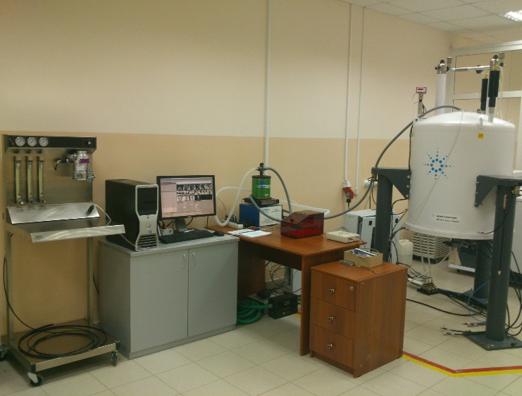
The Laboratory of Magnetic Resonance Imaging of Lobachevsky State University of Nizhni Novgorod is equipped with a new powerful MR tomograph for preclinical studies of pharmaceuticals. It has been successfully tested and started its work. The most important part of the experimental installation AgilentDDR2 400WB is a superconducting wide bore magnet that is much more powerful than usually used in hospitals - 9.4T compared with 1.5T.
On June 16, the team of the Laboratory of Magnetic Resonance Imaging of Lobachevsky State University of Nizhni Novgorod obtained the first results of tests of pharmaceuticals. PhD students, Alexander Romanov and Maria Muravyeva, and Associate Professor in the Department of Oncology at Oxford University Alexandr Khrapichev were supervised by Professor Irina Mukhina who is a head of Lobachevsky State University’s Biotechnology Development Centre - Research Institute known as the Institute of Living Systems.
The report says: “As the preliminary results, we are demonstrating here the first ever in vivo MR images taken in Nizhny Novgorod. There was preclinical test of morphological evidence of neuroprotective effect of potential pharmacological drug Carbamylated Erythropoietin (C-darbe) in the model of local ischemia or stroke due to photothrombosis in motor region of cerebral hemisphere. We used different regimes to create a set of MR images: the gradient echo (T2*-weighted) MR images, this technique is typically used for functional imaging to investigate dynamic process; anatomical (T2-weighted) MR images with the contrast proportional to the water content, and diffusion weighted MR images with the contrast inversely proportional to the diffusion”.
The successful results of tests were expected by the Laboratory of Magnetic Resonance Imaging of Lobachevsky State University of Nizhni Novgorod and its Oxford collaborator. This cooperation has already led to good outcomes with the efficiency of methods of magnetic resonance imaging raised and the formulae of potential pharmaceuticals improved. They will be all tested by experimental installation AgilentDDR2 400WB at Lobachevsky State University of Nizhni Novgorod.
Experts: Preclinical in vivo MR Imaging using the animal model
Magnetic Resonance Imaging (MRI) is one of the most promising techniques for in vivo imaging of alive bioobject due to non-invasiveness and absence of the harmful radiation. The image contrast is based on parameters specific to MRI such as T1 and T2 as well as on biological properties of tissue such as water content, blood flow and diffusion, etc. Another option is to use specific contrast agents to highlight the region of interest.
The main directions of research are the functional imaging (fMRI), i.e. measurement of temporal activity dependence changes of blood flow in the different parts of the brain; the diffusion tensor imaging (DTI), i.e. measurement of localized diffusion anisotropy to determine the microstructure of the brain; and the angiography (ASL) as mapping of blood flow distribution in the brain. In addition, we are going to use one of the most advanced MR techniques such as the chemical exchange saturation transfer (CEST) – enchantment of the contrast based on the magnetization transfer of highly diluted contrast agent weak signal to strong water signal; the latter can be registered directly.
The NMRI laboratory of Lobachevsky State University of Nizhni Novgorod is equipped with 9.4T superconducting wide bore magnet driven by Agilent DDR2 console.
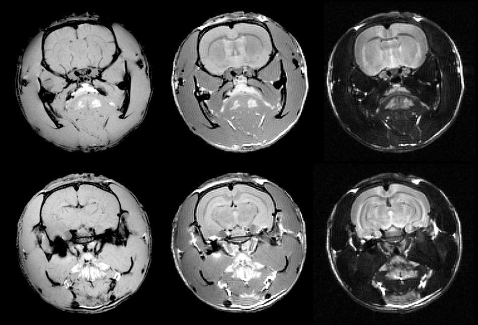
This figure shows an example of MR images of an intact rat brain and rat brain with local ischemia.


