
New implant architecture for accelerated bone tissue regeneration proposed by Nizhny Novgorod scientists
Scientists from Nizhny Novgorod have come up with a solution for the rapid regeneration of large bone defects. Experiments on laboratory animals have already shown that bone regeneration with heterogeneous implants is twice as fast as with standard implants having a homogeneous structure.
The project is implemented by biomedical researchers at the Institute of Clinical Medicine of Lobachevsky University (Nizhny Novgorod), Privolzhsky Medical Research University (Nizhny Novgorod), and scientists at Sechenov University (Moscow), Semenov Institute of Chemical Physics, Russian Academy of Sciences (Moscow) and the Crystallography and Photonics Research Centre, Russian Academy of Sciences (Moscow).
Even using the patient's own bone does not guarantee complete closure of the defect, and bone material from another person may not engraft at all. The main goal of the project is to obtain synthetic implants with characteristics as close as possible to those of human bone. A bone model is created using 3D laser printing, its scaffold cells are seeded with the patient's stem cells, and the entire structure is implanted into the damaged area.
"The synthetic bone tissue consists of multiple cells that replicate the heterogeneous structure of bone: with large pores inside the implant and smaller ones on the surface. This structure allows blood vessels to proliferate, and the rate of scaffold biodegradation corresponds to the rate of bone repair," explained Daria Kuznetsova, Head of the Laboratory for Molecular Genetic Research at the UNN Institute of Clinical Medicine.
In their research, the scientists recreated a fragment of a mouse skull represented by nanoscaffolds with a three-layer structure. The implants made of biocompatible lactic acid-based material were printed on a laser 3D printer at Sechenov University. The technology makes it possible to adjust the rate of biodegradation of the material so that the scaffold is decomposed as the bone recovers.
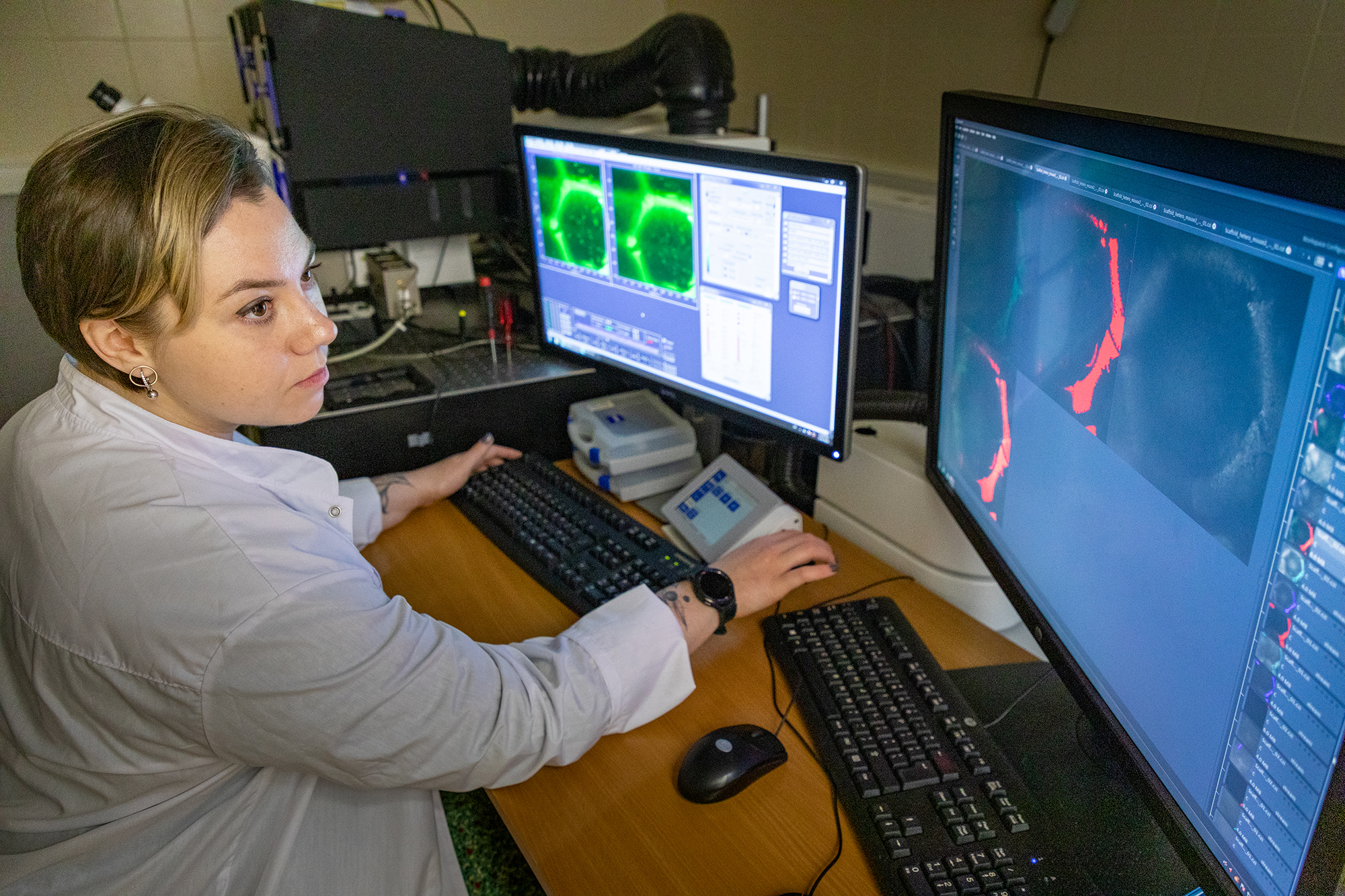
"Bone regeneration was shown by metabolic imaging and the biodegradation of the implant was assessed by fluorescence microscopy. Through a combination of these imaging techniques, we proved that the delivery of oxygen, nutrients, mineralisation and other processes are more intensive on heterogeneous scaffolds," said Daria Kuznetsova.
In the future, the authors plan to improve the composition of the synthetic bone tissue and increase the size of the scaffolds. The research was carried out as part of the federal programme "Priority 2030". The results are published in the international biomedical scientific journal Stem Cell Research & Therapy.


